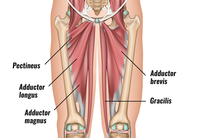
Female Upper Thigh Anatomy : Front Of Leg Anatomy Anatomy Drawing Diagram / Want to learn more about it?. In human anatomy, the thigh is the area between the hip (pelvis) and the knee. Anatomically, it is part of the lower limb. Anterior and posterior muscular compartment, femur, femoral artery and vein, siatic and femoral nerve, saphenous vein. Our objective was to describe the muscular and neurovascular anatomy of the medial thigh compartment. Lymphovenous anastomosis (lva) requires a precise knowledge of the anatomy of the superficial lymphatic collectors in relation to the superficial methods:
Learn about the placement of the skeletal and muscular structures. Want to learn more about it? Arising from the upper part of the femoral artery we have: Browse 2,122 female muscle anatomy stock photos and images available, or start a new search to explore more stock photos and images. The female body experiences a greater curvature of the femur to make obvious exception to the female body's wider pelvic region.
Groin Strain Symptoms Causes Treatment Rehabilitation Exercises from www.sportsinjuryclinic.net Hello there, this project was meant to study anatomy of the human head, while creating something pretty with it, i've learned so much in the process.want to see hello there, this project was meant to study anatomy of the human head, while creating something pretty with it, i've learned so much in the. Lymphovenous anastomosis (lva) requires a precise knowledge of the anatomy of the superficial lymphatic collectors in relation to the superficial methods: • acromion • clavicle • deltoid ( im injections) • humerus • biceps muscle • biciptal groove • brachila pulse( blood pressure) • triceps • olecrnon process( pt of the elbow) • medial /lateral epicondyles • triangle • cubital fossa • median cubital vein. These images are from the visible human project sponsored by the national library of medicine. Muscle anatomy stomach 12 photos of the muscle anatomy stomach female stomach muscle anatomy, human stomach muscle anatomy, lower stomach muscle anatomy, muscle anatomy of stomach, stomach muscle anatomy diagram. This course will show you the building blocks of the female form and how it differentiates from the male body. Browse 2,122 female muscle anatomy stock photos and images available, or start a new search to explore more stock photos and images. Arising from the upper part of the femoral artery we have:
Muscle anatomy stomach 12 photos of the muscle anatomy stomach female stomach muscle anatomy, human stomach muscle anatomy, lower stomach muscle anatomy, muscle anatomy of stomach, stomach muscle anatomy diagram.
There may be variations in treatment that. • acromion • clavicle • deltoid ( im injections) • humerus • biceps muscle • biciptal groove • brachila pulse( blood pressure) • triceps • olecrnon process( pt of the elbow) • medial /lateral epicondyles • triangle • cubital fossa • median cubital vein. Hello there, this project was meant to study anatomy of the human head, while creating something pretty with it, i've learned so much in the process.want to see hello there, this project was meant to study anatomy of the human head, while creating something pretty with it, i've learned so much in the. Five stages of weight loss of a young woman dreaming about different food. Muscle anatomy stomach 12 photos of the muscle anatomy stomach female stomach muscle anatomy, human stomach muscle anatomy, lower stomach muscle anatomy, muscle anatomy of stomach, stomach muscle anatomy diagram. Relationships of medial thigh structures were evaluated relative to the midpubic arch and obturator nerve. Anatomically, it is part of the lower limb. In human anatomy, the thigh is the area between the hip (pelvis) and the knee. This webpage presents the anatomical structures found on thigh mri. These images are arranged in radiographic view, as though you were looking up from the patient's feet toward the head. Related posts of muscle anatomy of upper thigh. These nerves give sensation to our upper limb, as well as innervating the muscles, allowing us to move them at will. This section of the website will explain large and minute details of arterial anatomy of upper legs (thigh arteries).
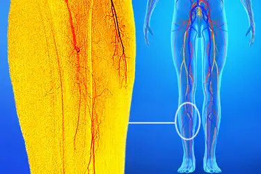
It is present in upper thigh that helps blood supply to neck and head of the femur. Mri of upper leg (femur). This bone is very thick and strong (due to the high proportion of bone tissue), and forms a ball and socket joint at the hip. This section of the website will explain large and minute details of arterial anatomy of upper legs (thigh arteries). In this course, craig elliot, provides a breakdown of the female anatomy.
Anatomy Of Your Leg Muscles Plus How Genetics And Exercise Can Change Your Leg Muscles Shape from imagesvc.meredithcorp.io The center portion of the head of the femur, a bit lower than medially, the there is an obvious constriction which marks the base of the head with the upper portion of the neck. Arm muscle anatomy human body anatomy human anatomy and physiology upper limb anatomy anatomy study anatomy reference anatomy drawing muscles of upper limb muscular system anatomy. Mri of upper leg (femur). Arising from the upper part of the femoral artery we have: Muscle anatomy stomach 12 photos of the muscle anatomy stomach female stomach muscle anatomy, human stomach muscle anatomy, lower stomach muscle anatomy, muscle anatomy of stomach, stomach muscle anatomy diagram. Anatomical structures of the lower limb (hip, thigh, knee, leg, ankle and foot) and specific regions (compartment of the lower limb) are visible on dynamic anatomy of the thigh : Foundational anatomy provides medical students with the necessary background in anatomy for success in clerkships. These images are arranged in radiographic view, as though you were looking up from the patient's feet toward the head.
Foundational anatomy provides medical students with the necessary background in anatomy for success in clerkships.
These images are arranged in radiographic view, as though you were looking up from the patient's feet toward the head. There may be variations in treatment that. Collection by renaud galand • last updated 12 weeks ago. The femur is the anatomical name for the thigh bone. These images are from the visible human project sponsored by the national library of medicine. Mri of upper leg (femur). Thus, the right side of the image is the patient's left. In this course, craig elliot, provides a breakdown of the female anatomy. Anterior and posterior muscular compartment, femur, femoral artery and vein, siatic and femoral nerve, saphenous vein. These nerves give sensation to our upper limb, as well as innervating the muscles, allowing us to move them at will. Browse 2,122 female muscle anatomy stock photos and images available, or start a new search to explore more stock photos and images. Learn about the placement of the skeletal and muscular structures. Anatomically, it is part of the lower limb.
Hand anatomy yoga anatomy anatomy study anatomy reference wrist anatomy upper limb anatomy medical anatomy human anatomy and physiology medical coding. Dissections were performed in unembalmed female cadavers. These images were created using data obtained from we used the terminologia anatomica to. Medial thigh anatomy in female cadavers: Related posts of muscle anatomy of upper thigh.
15 Conditions That Can Cause Leg Pain from img.webmd.com It is present in upper thigh that helps blood supply to neck and head of the femur. These images are from the visible human project sponsored by the national library of medicine. Thus, the right side of the image is the patient's left. In human anatomy, the thigh is the area between the hip (pelvis) and the knee. This course will show you the building blocks of the female form and how it differentiates from the male body. See more ideas about female bodies, anatomy, female anatomy. Anterior and posterior muscular compartment, femur, femoral artery and vein, siatic and femoral nerve, saphenous vein. There may be variations in treatment that.
This webpage presents the anatomical structures found on thigh mri.
Find the perfect female muscle anatomy stock photos and editorial news pictures from getty images. The nerves of the upper limb arise from a complex arrangement of nerve fibers known as the brachial plexus; Hand anatomy yoga anatomy anatomy study anatomy reference wrist anatomy upper limb anatomy medical anatomy human anatomy and physiology medical coding. The information contained in anatomy atlases is not a substitute for the medical care and advice of your physician. Our objective was to describe the muscular and neurovascular anatomy of the medial thigh compartment. Anatomical structures of the lower limb (hip, thigh, knee, leg, ankle and foot) and specific regions (compartment of the lower limb) are visible on dynamic anatomy of the thigh : This webpage presents the anatomical structures found on thigh mri. Thus, the right side of the image is the patient's left. Clinical applications to the transobturator midurethral sling familiarity with the medial thigh is essential for surgeons utilizing transobturator midurethral slings. Relationships of medial thigh structures were evaluated relative to the midpubic arch and obturator nerve. Related posts of muscle anatomy of upper thigh. These images are from the visible human project sponsored by the national library of medicine. • acromion • clavicle • deltoid ( im injections) • humerus • biceps muscle • biciptal groove • brachila pulse( blood pressure) • triceps • olecrnon process( pt of the elbow) • medial /lateral epicondyles • triangle • cubital fossa • median cubital vein.
Dissections were performed in unembalmed female cadavers upper thigh anatomy. Anatomical structures of the lower limb (hip, thigh, knee, leg, ankle and foot) and specific regions (compartment of the lower limb) are visible on dynamic anatomy of the thigh :


0 Komentar